MPFL RECONSTRUCTION
What is MPFL reconstruction?
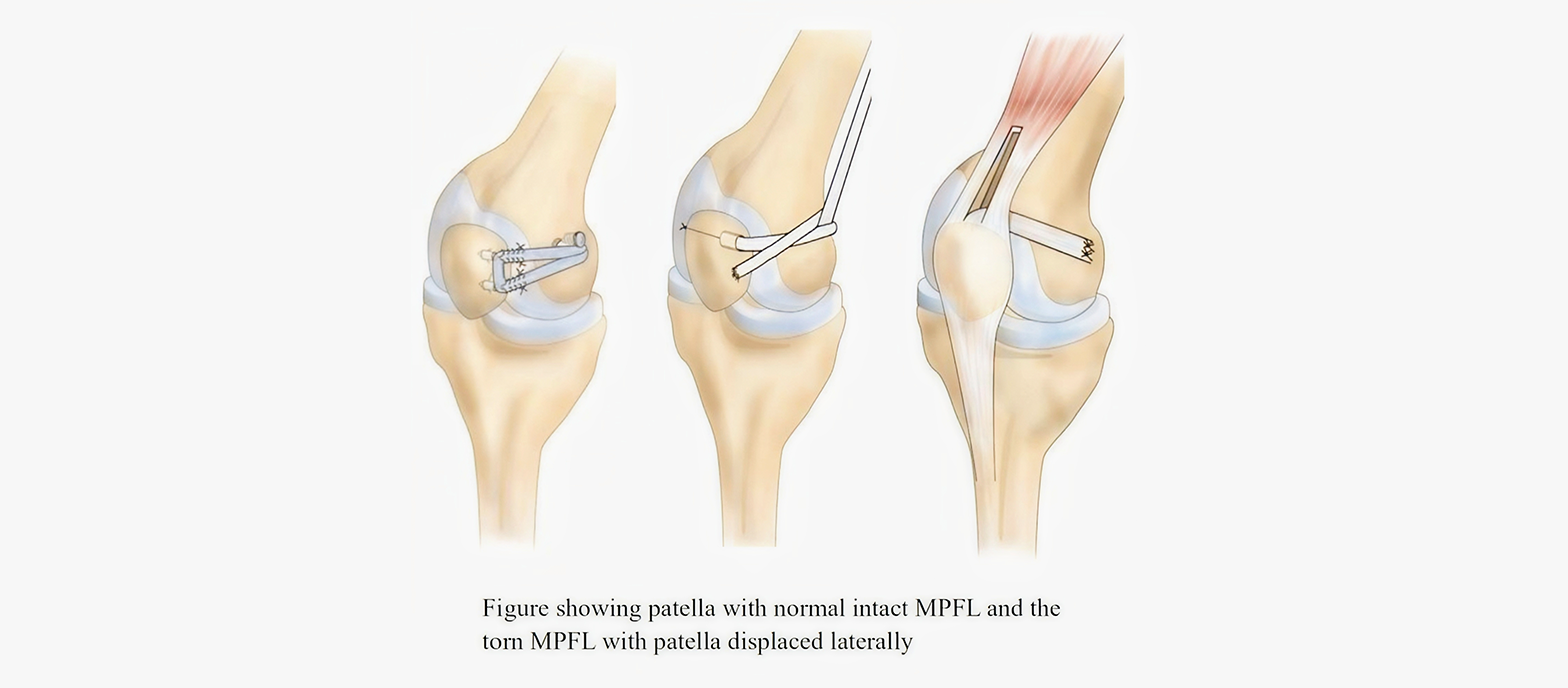
The MPFL (Medial Patellofemoral Ligament) is a band of tissue on the inner side of the knee. It helpsto keep the kneecap (patella) in place and stops it from sliding out of position.
When does the MPFL get injured?
The MPFL can get torn if your kneecap slips out (dislocates), usually due to a sudden twist, fall, or sporting activity.
Who will get MPFL injury?
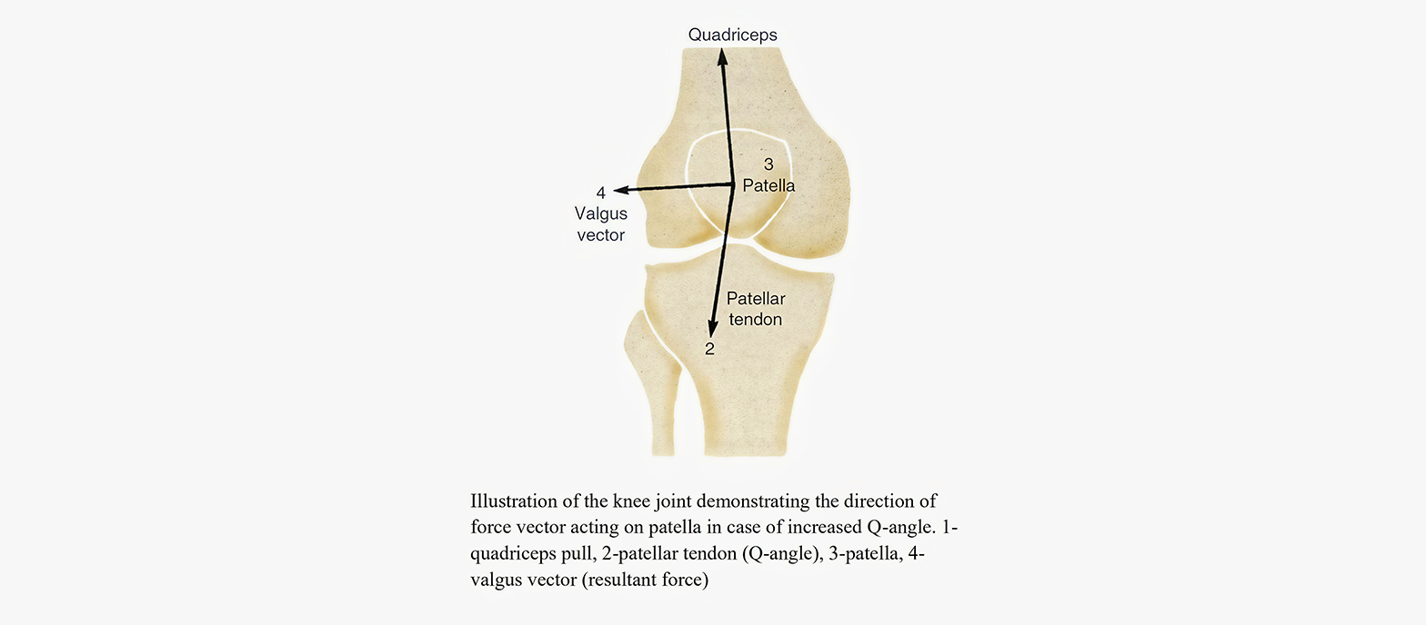
Though it mostly occur due to trauma in the adults, MPFL injury has a prepondence to those who have a slightly different shape of thigh bone either at the hip or at the knee. The leg bone (tibia), muscles and l,igaments next to the knee also play a role in the slipping of knee cap.
What are the symptoms?
If the MPFL is torn, your kneecap may feel loose or unstable. You might feel pain in the front and inner side of your knee, and it ay feel like the kneecap is slipping during movement.
What is MPFL reconstruction?
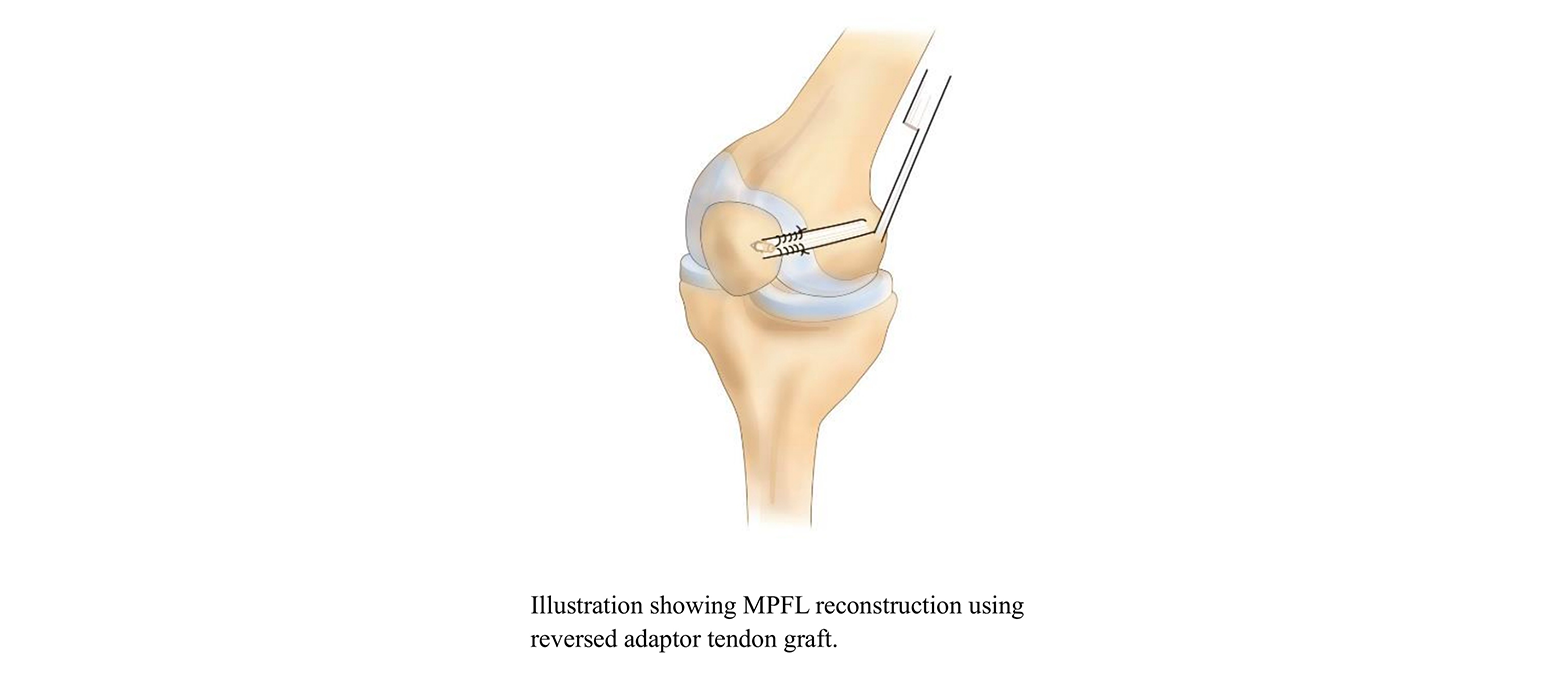
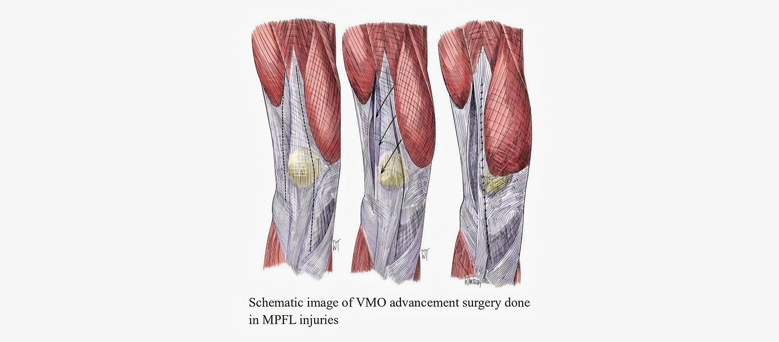
It is a surgery to fix or replace the torn MPFL. The doctor uses a tendon graft (from your body or a donor) to reconstruct the ligament and realign your patella.
What are the surgical procedures?
The surgeon makes small cuts near your kneecap. A freshly harvested tendon (usually from your harmstring) is attached on the side of the patella and the thigh bone to realign in the centre.
How soon can I recover?
You’ll wear a knee brace after surgery. Dr. Sudhan will give you an individualised protocol based on your problem and the treatment given. Which is usually starts with regaining movements and steps up to strengthening. Physical rehabilitation is the key. Most people recover fully in 4 to 6 months.
Are there any risks?
No specific risk is associated with MPFL reconstruction, and it is generally considered safe. The front of your knee will have an incision scar, which may be variable from 2.5cm to 5 cm in length.
MEDIAL PLICATION AND LATERAL RELEASE PROCEDURE
What is this procedure for?
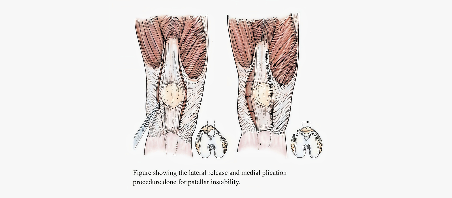
This procedure is done to help correct the kneecap that does not stay in place or dislocates either in daily activities or during sports activities.
What is Medial Plication?
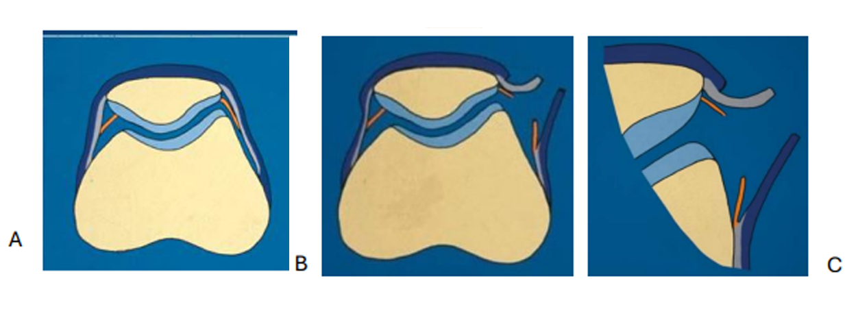
Medial plication means tightening the inner side of the kneecap. The doctor stitches or folds the soft tissues on the inside of the knee to pull the kneecap back toward the centre.
What is Lateral Release?
Lateral release means loosening the outer side of the kneecap. The doctor cuts the tight tissues on the outside of the knee to let the kneecap move more freely into its normal place.
How is the procedure done?
The procedure is usually done using a small camera (arthroscope). Tiny cuts are made around the knee, and appropriate instruments are used to do the tightening and loosening as needed. It is a minimally invasive surgery.
Are there any risks with the procedure?
No specific risk is associated with the procedure. Generally, it is considered safe.
TIBIALTUBERCLE TRANSFER
What is tibial tubercle transfer?
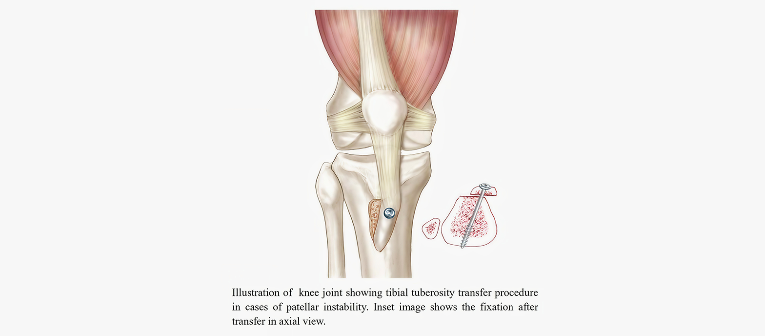
Tibial tubercle transfer is a surgery that helps fix the position of the kneecap (patella) when it moves out of place, often during physical activity. The surgeon cuts a small part of the shin bone (tibial tubercle), which is where the patellar tendon attaches, and moves it to a better position. This prevents the kneecap from dislocation and helps move properly, and reduces discomfort.
What is the Elmslie-Trillat operation?
It is a type of tibial tuberosity transfer procedure done to treat an unstable kneecap that dislocates often and easily. During the surgery, the tibial tubercle is shifted towards the inner side of the knee and fixed using screws, with or without medial plication & lateral release (modified Elmslie-Trillat). This helps the kneecap stay in the correct path and prevents it from slipping out. It is often used for people who frequently experience kneecap dislocation.
What is Fulkerson Osteotomy?
Fulkerson Osteotomy is another type of surgery used to treat kneecap alignment issues. In this procedure, the tibial tubercle is moved both forward and towards the inside. This change reduces the pressure between the kneecap and the thigh bone (femur) and also helps prevent dislocation. It is helpful for people who have pain due to the kneecap rubbing on the thigh bone.
Why are these procedures done?
These procedures are done to correct the alignment of the kneecap, especially when the kneecap and the tibia (shin bone) are not aligned properly. The goal is to improve the movement and position of the kneecap.
What are the common symptoms?
People who need these surgeries are the one who has kneecap alignment issues which causes pain in the front of the knee, kneecap slipping while walking or running, clicking and grinding sounds on movement. These persons may also find it hard to use stairs or squat comfortably
How is the condition diagnosed?
To diagnose the condition, the doctor will ask about your knee symptoms and check how the kneecap moves, and conduct some special tests. They may also take X-rays or an MRI scan to see the alignment of the bones and check for damage to the cartilage.